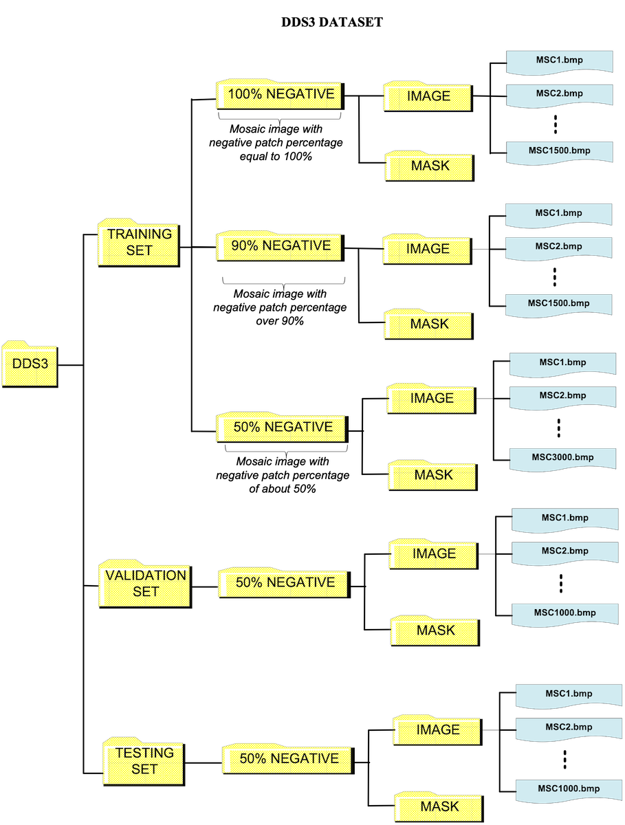
The DDS3 Dataset consists of 8,000 mosaic images with a size of 400x400 pixels and their respective masks.
Mosaic images were generated with positive and negative patches taken from the digital fields of slides S01 to S15. Masks consists of a binary image with bacilli pixels inwhite and background pixels black.
The training set is composed of 6000 images, with 3000 mosaic images with about 50% positive and 50% negative patches, 1500 mosaic images with over 90% negative patches, and 1500 mosaic images with 100% negative patches.
The validation set is composed of 1000 mosaic images with about 50% positive and 50% negative patches.
The testing set is composed of 1000 mosaic images with about 50% positive and 50% negative patches. The flowchart below shows the organization of the data.
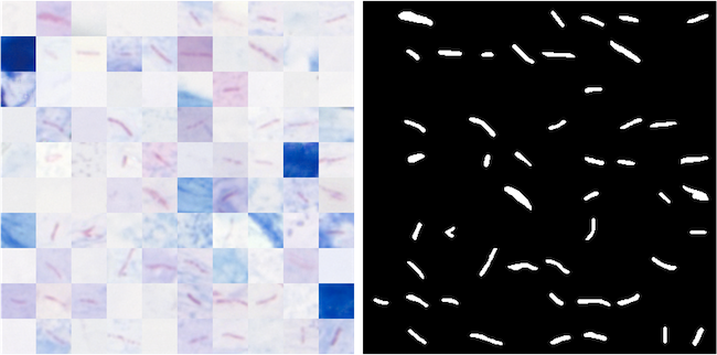
Figure 1. Example of mosaic image with 400x400 pixels and respective binary mask.
ABBREVIATIONS, NOMENCLATURE AND FILE DIRECTORY
ABBREVIATIONS
S → Slide
T → Tile (a digital microscopy field)
TR →Tile Region (a set of 164 Tiles or digital fields)
St → Stack (a set of 11 digital images acquired from a microscopic field at different focal depths)
EDF→ Extended Depth of Focus image (a digital image obtained by the fusion of the stack of 11 images)
M → Marking (Annotation)
NP → Negative Patch
PP→ Positive Patch
MSC→ Mosaic Image